Medical Image Segmentation: GI Tract
🏥 Clinical Motivation
Radiation therapy for gastrointestinal tract cancers demands sub-millimeter precision. The stomach and intestines shift position daily responding to digestion, breathing, and organ filling what makes static treatment plans dangerous.
While modern MR-Linac systems acquire real-time MRI scans during radiation delivery, manually segmenting these anatomical structures before each session is incredibly labor-intensive and introduces inter-observer variability.
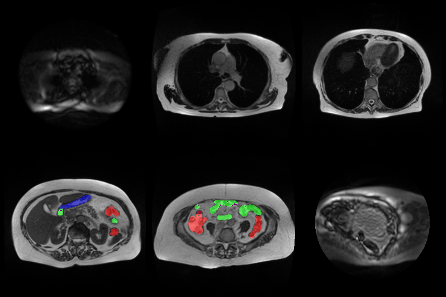
The Solution: This project automates the segmentation process. Given an MRI scan, the model simultaneously and accurately delineates three critical structures in real-time:
- 🔴 Large Bowel (Red Mask)
- 🟢 Small Bowel (Green Mask)
- 🔵 Stomach (Blue Mask)
This automation enables oncologists to adapt radiation beams to daily organ positions, heavily targeting tumors while strictly sparing healthy tissue.
🎥 Project Demo
📊 Dataset & Pipeline
Trained on the UW-Madison GI Tract Image Segmentation clinical dataset, featuring high intra-patient variability, varying contrast, and thin-slice MRI scans.
Custom Data Pipeline (dataset.py):
- Decodes complex Run-Length Encoded (RLE) masks on-the-fly into binary segmentation maps.
- Stacks masks into 3-channel tensors (one channel per target organ).
- Implements robust data augmentation and memory-efficient loading with RAM caching capabilities.
🧠 Benchmarked Architectures
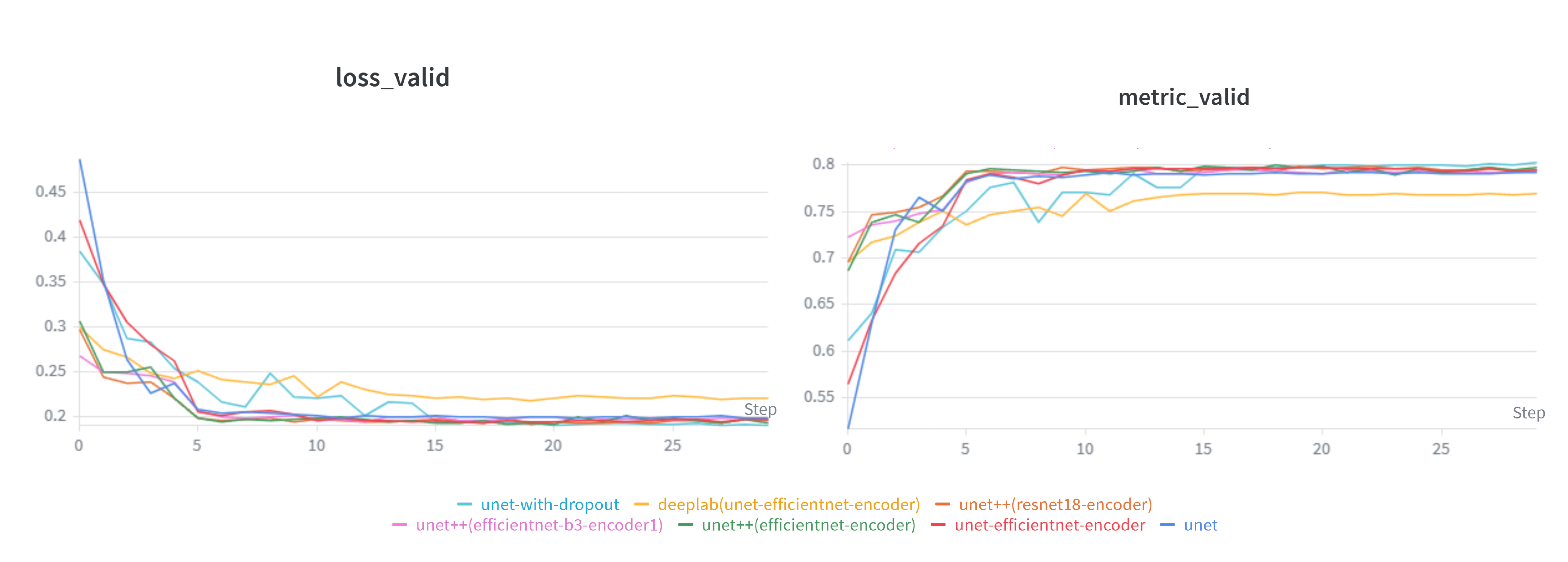
This repository moves beyond a single model, offering a systematic benchmark of multiple segmentation topologies:
1. Custom U-Net (From Scratch)
A pure PyTorch implementation featuring successive 3x3 convolution blocks, a deep context bottleneck, and transposed convolution upsampling with skip connections to preserve fine spatial details.
2. U-Net with Pre-trained Encoders
Utilizes transfer learning by replacing the standard encoder with ImageNet-pretrained backbones:
- EfficientNet-B4: Delivers the highest accuracy-to-FLOP ratio.
- ResNet-50 / ResNet-101: Provides exceptionally stable feature hierarchies.
- SE-ResNeXt: Integrates Squeeze-and-Excitation blocks for channel-wise attention, boosting sensitivity to subtle, ambiguous organ boundaries.
3. U-Net++
A nested, densely connected architecture that bridges the semantic gap between encoder and decoder feature maps. Intermediate nodes aggregate features progressively, heavily improving performance on complex, varying shapes.
4. DeepLabV3+
Leverages Atrous Spatial Pyramid Pooling (ASPP). By using dilated convolutions, it expands the receptive field to capture multi-scale context without downsampling—proving highly effective for irregular, non-convex structures like the small bowel.
🔬 Loss Functions & Optimization
Medical datasets suffer from severe class imbalance (background pixels vastly outnumber organ pixels). To counter this, the loss/ module implements a highly modular suite:
- Binary Cross-Entropy (BCE): The standard pixel-wise baseline.
- Dice Loss: Directly optimizes the volumetric overlap, completely bypassing background imbalance.
- BCE + Dice Hybrid: The optimal standard—BCE stabilizes gradients while Dice forces boundary accuracy.
- Focal & Tversky Loss: Advanced formulations to down-weight easy negatives and control the recall-precision tradeoff.
🛠️ Infrastructure & Inference
- Inference Engine (
segment.py): A streamlined script that loads models, processes raw scans, and generates side-by-side color-coded overlays (mirroring clinical annotation standards) for intuitive visual evaluation. - Experiment Runner (
Experiments.py): Automates multi-model and multi-loss training runs with automatic result logging. - Hyperparameter Sweeps (
HP.py): Systematic grid-search architecture over learning rates, weight decays, and loss combinations.